39 light microscope with labels
Electron microscope - Wikipedia An electron microscope is a microscope that uses a beam of accelerated electrons as a source of illumination. As the wavelength of an electron can be up to 100,000 times shorter than that of visible light photons, electron microscopes have a higher resolving power than light microscopes and can reveal the structure of smaller objects. Light Microscope Parts, Function & Uses - Study.com A light microscope definition is an instrument that uses light, magnifying lenses, and an eyepiece to examine objects too small to be seen by the naked eye. Light microscopes have several uses.
Parts of a microscope with functions and labeled diagram - Microbe Notes Microscopic illuminator - This is the microscopes light source, located at the base. It is used instead of a mirror. It captures light from an external source of a low voltage of about 100v. Condenser - These are lenses that are used to collect and focus light from the illuminator into the specimen.

Light microscope with labels
ProSciTech Laboratory supplies and Lab equipment for Histology, Pathology, Light Microscopy, Electron Microscopy and specialist researchers. Microscope Objective Lens | Products | Leica Microsystems The objective lens is a critical part of the microscope optics. The microscope objective is positioned near the sample, specimen, or object being observed. It has a very important role in imaging, as it forms the first magnified image of the sample. The numerical aperture (NA) of the objective indicates its ability to gather light and largely determines the microscope’s resolution, the ... Parts of the Microscope with Labeling (also Free Printouts) Parts of the Microscope with Labeling (also Free Printouts) By Editorial Team March 7, 2022 A microscope is one of the invaluable tools in the laboratory setting. It is used to observe things that cannot be seen by the naked eye. Table of Contents 1. Eyepiece 2. Body tube/Head 3. Turret/Nose piece 4. Objective lenses 5. Knobs (fine and coarse) 6.
Light microscope with labels. Compound Microscope Labeled Diagram | Quizlet QUESTION. The total magnification of a specimen being viewed with a 10X ocular lens and a 40X objective lens is. 15 answers. QUESTION. a mosquito beats its wings up and down 600 times per second, which you hear as a very annoying 600 Hz sound. if the air outside is 20 C, how far would a sound wave travel between wing beats. 2 answers. Labeling the Parts of the Microscope | Microscope World Resources Labeling the Parts of the Microscope This activity has been designed for use in homes and schools. Each microscope layout (both blank and the version with answers) are available as PDF downloads. You can view a more in-depth review of each part of the microscope here. Download the Label the Parts of the Microscope PDF printable version here. Compound Light Microscope: Everything You Need to Know A fluorescence microscope, also called a confocal microscope, is a kind of biological microscope that operates by using different light colors and wavelengths over-dyed specimen samples in order for the dye to interact with the light, after which the resulting image is scanned. Label the microscope — Science Learning Hub All microscopes share features in common. In this interactive, you can label the different parts of a microscope. Use this with the Microscope parts activity to help students identify and label the main parts of a microscope and then describe their functions. Drag and drop the text labels onto the microscope diagram.
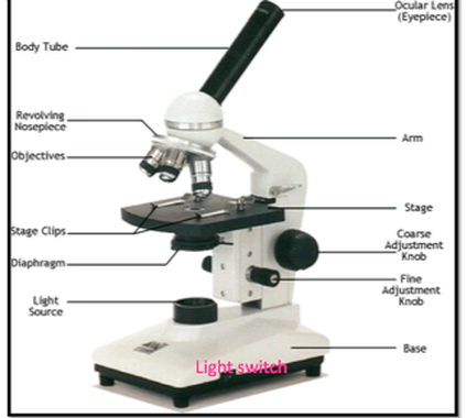
A Study of the Microscope and its Functions With a Labeled Diagram ... The microscope is an important instrument in the world of biological science. Diagrams have always been of great help in understanding both the structural and functional aspects of entities. These labeled microscope diagrams and the functions of its various parts, attempt to simplify the microscope for you. PDF Parts of the Light Microscope - Science Spot Supports the MICROSCOPE D. STAGE CLIPS HOLD the slide in place C. OBJECTIVE LENSES Magnification ranges from 10 X to 40 X F. LIGHT SOURCE Projects light UPWARDS through the diaphragm, the SPECIMEN, and the LENSES H. DIAPHRAGM Regulates the amount of LIGHT on the specimen E. STAGE Supports the SLIDE being viewed K. ARM Used to SUPPORT the Microscope Labeling - The Biology Corner 1) Start with scanning (the shortest objective) and only use the COARSE knob . Once it is focused… 2) Switch to low power (medium) and only use the COARSE knob . You may need to recenter your slide. Once it is focused.. 3) Switch to high power (long objective). Microscope, Microscope Parts, Labeled Diagram, and Functions Revolving Nosepiece or Turret: Turret is the part of the microscope that holds two or multiple objective lenses and helps to rotate objective lenses and also helps to easily change power. Objective Lenses: Three are 3 or 4 objective lenses on a microscope. The objective lenses almost always consist of 4x, 10x, 40x and 100x powers. The most common eyepiece lens is 10x and when it coupled with ...
Microscope Types (with labeled diagrams) and Functions This is an advanced microscope that has specific application in viewing, observing and measuring the optical thickness and phase of completely transparent specimens and objects. A tiny interferometer is used and a specimen is placed on beam path of it. This path is split and then rejoined to create two superimposed images of the specimen in focus. Label the Light Microscope - Labelled diagram - Wordwall Drag and drop the pins to their correct place on the image.. Eyepiece, Light Source, Base, Stage, Stage Clips, Fine Focus, Coarse Focus, Arm, Objective Lens. Light Microscope: Functions, Parts and How to Use It To use a light microscope, you can follow the steps below carefully. Start with a low lens and a clean slide. The microscope stage should be lowered as low as possible. Center the slide so that the specimen is under the objective lens. Use the coarse adjustment knob to get a general focus. Then slowly move up the stage until focus is achieved. ZEISS Lightsheet 7 – Light Sheet Microscope This effect occurs in all fluorescence microscopes, but the illumination axis in light sheet fluorescence microscopy is perpendicular to the observation axis and so this effect is more obvious. In Lightsheet 7, a patented Pivot Scanner alters the angle of the light sheet upwards and downwards during image acquisition.
A Light Microscope - Micropedia
What is label in microscope? - Gowanusballroom.com Parts of the Microscope with Labeling (also Free Printouts) 1. Eyepiece. Through the eyepiece, you can visualize the object being studied. Its magnification capacity ranges between… 2. Body tube/Head. It is the structure that connects the eyepiece to the lenses. Image 2: The body tube part of a… Is there a print version of the microscope layout?

Neuron cell under a microscope | Science and Discovery | Pinterest | Neurons
Student's Guide: How to Use a Light Microscope An ultraviolet microscope uses UV light to view specimens at a resolution that isn't possible with the common brightfield microscope. It utilizes UV optics, light sources, as well as cameras. Because of the shorter wavelengths of UV light (180-400 nm), the image produced is clearer and more distinct at a magnification approximately double what ...
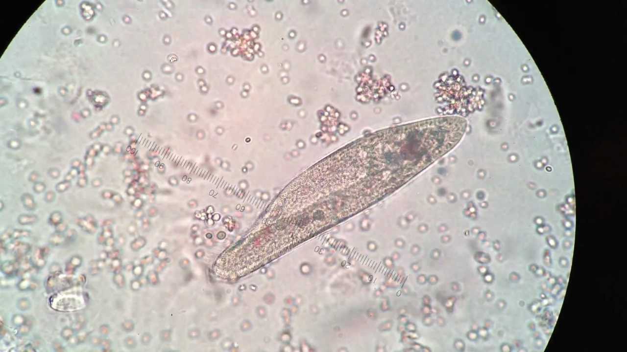
Paramecium under 400X magnification - YouTube
Light Microscope- Definition, Principle, Types, Parts, Labeled Diagram ... A light microscope is a biology laboratory instrument or tool, that uses visible light to detect and magnify very small objects and enlarge them. They use lenses to focus light on the specimen, magnifying it thus producing an image. The specimen is normally placed close to the microscopic lens.

Unlabeled Compound Light Microscope Diagram - Micropedia
Compound Microscope Parts, Functions, and Labeled Diagram Compound Microscope Definitions for Labels. Eyepiece (ocular lens) with or without Pointer: The part that is looked through at the top of the compound microscope. Eyepieces typically have a magnification between 5x & 30x. Monocular or Binocular Head: Structural support that holds & connects the eyepieces to the objective lenses.
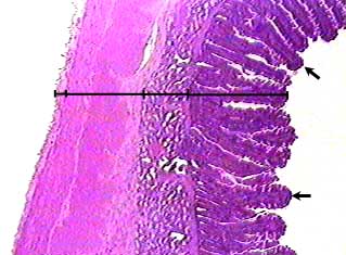
Duodenum
Microscope Parts and Functions Microscope Parts and Functions With Labeled Diagram and Functions How does a Compound Microscope Work?. Before exploring microscope parts and functions, you should probably understand that the compound light microscope is more complicated than just a microscope with more than one lens.. First, the purpose of a microscope is to magnify a small object or to magnify the fine details of a larger ...
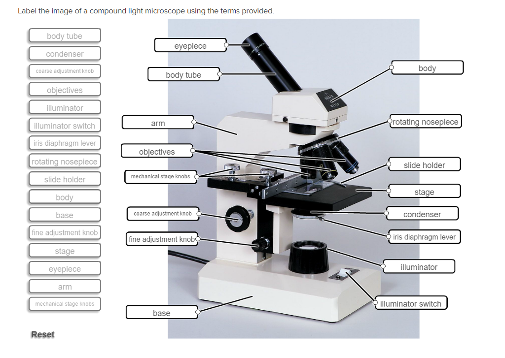
Solved: Label The Image Of A Compound Light Microscope Usi... | Chegg.com
Light microscopes - Cell structure - Edexcel - BBC Bitesize Microscopes are used to produce magnified images. There are two main types of microscope: light microscopes are used to study living cells and for regular use when relatively low magnification and...
Using the Microscope
Sperm Under Microscope with Labeled Diagram - AnatomyLearner Under the light microscope, the sperm consists of two main portions - the head and the tail. But, the electron microscope shows four different parts in the tail of spermatozoa. ... So, this article provides the details structural features of sperm under the light microscope. All the labeled diagrams might help you identify the sperms from ...
Using a Light Microscope - AyushiSinhaMicroscopy
ZEISS Axioscope 5 Smart Laboratory Microscope Focus. Snap. Done. Forget about the 15 steps and clicks to document samples with multiple fluorescent labels. With Smart Microscopy, this is a thing of the past. Axioscope 5 with Axiocam 202 mono and Colibri 3 LED illumination take this workload from you. You keep your hands at the microscope stand. Relaxed.

Types Of Light Microscopes
Microscope Labeling Game - PurposeGames.com This is an online quiz called Microscope Labeling Game There is a printable worksheet available for download here so you can take the quiz with pen and paper. This quiz has tags. Click on the tags below to find other quizzes on the same subject. Science microsope Total Points 0 Get started! Today's Rank -- 0 's Points Points 15

Zellkern Dr.Jastrows EM-Atlas
Parts of Stereo Microscope (Dissecting microscope) – labeled ... Labeled part diagram of a stereo microscope Major structural parts of a stereo microscope. There are three major structural parts of a stereo microscope. The viewing Head includes the upper part of the microscope, which houses the most critical optical components, including the eyepiece, objective lens, and light source of the microscope.

BIO 156, Fall 2015: Week 8. Lab 7. Immune System
Light Microscope Labeled - how scanning electron microscopes work ... Light Microscope Labeled - 16 images - senior biology cell theory microscopy, what is a light microscope with pictures, 29 you will love labeling a compound microscope db, microscope imaging station gallery,
302 Found
An Introduction to the Light Microscope, Light Microscopy Techniques ... For example, using green light with a wavelength of 550 nm and an objective with a typical NA of 0.7, a standard light microscope can resolve features down to a limit of 0.61 × ... Examples include biological samples that are intrinsically fluorescent or have been labeled with a fluorescent marker, as well as single molecules and other ...
Label a microscope - Teaching resources
Compound Microscope Parts - Labeled Diagram and their Functions The eyepiece (or ocular lens) is the lens part at the top of a microscope that the viewer looks through. The standard eyepiece has a magnification of 10x. You may exchange with an optional eyepiece ranging from 5x - 30x. [In this figure] The structure inside an eyepiece. The current design of the eyepiece is no longer a single convex lens.

A Light Microscope - Micropedia
Labelled Diagram Of A Light Microscope | Products & Suppliers ... Products/Services for Labelled Diagram Of A Light Microscope Microscopes - (706 companies) ...and transmission electron microscopes. Acoustic and ultrasonic microscopes use sound waves to create images of the sample. Compound microscopes use a single light path. These types of microscopes can have a single eyepiece (monocular) or a dual eyepiece...

Euglena Acus 2 - BF microscope 1250x - YouTube
Compound Microscope - Diagram (Parts labelled), Principle and Uses Also called as binocular microscope or compound light microscope, it is a remarkable magnification tool that employs a combination of lenses to magnify the image of a sample that is not visible to the naked eye. Compound microscopes find most use in cases where the magnification required is of the higher order (40 - 1000x).

compound light microscope labeled pp yyolwqzifkcwpieoo7qpy1b9flnvylgukhbeej7nfy ...
Parts of the Microscope with Labeling (also Free Printouts) Parts of the Microscope with Labeling (also Free Printouts) By Editorial Team March 7, 2022 A microscope is one of the invaluable tools in the laboratory setting. It is used to observe things that cannot be seen by the naked eye. Table of Contents 1. Eyepiece 2. Body tube/Head 3. Turret/Nose piece 4. Objective lenses 5. Knobs (fine and coarse) 6.

Blood and Blood Types | Under The Microscope - YouTube
Microscope Objective Lens | Products | Leica Microsystems The objective lens is a critical part of the microscope optics. The microscope objective is positioned near the sample, specimen, or object being observed. It has a very important role in imaging, as it forms the first magnified image of the sample. The numerical aperture (NA) of the objective indicates its ability to gather light and largely determines the microscope’s resolution, the ...

Urinary Bladder
ProSciTech Laboratory supplies and Lab equipment for Histology, Pathology, Light Microscopy, Electron Microscopy and specialist researchers.
Post a Comment for "39 light microscope with labels"