43 easy microscope diagram with labels
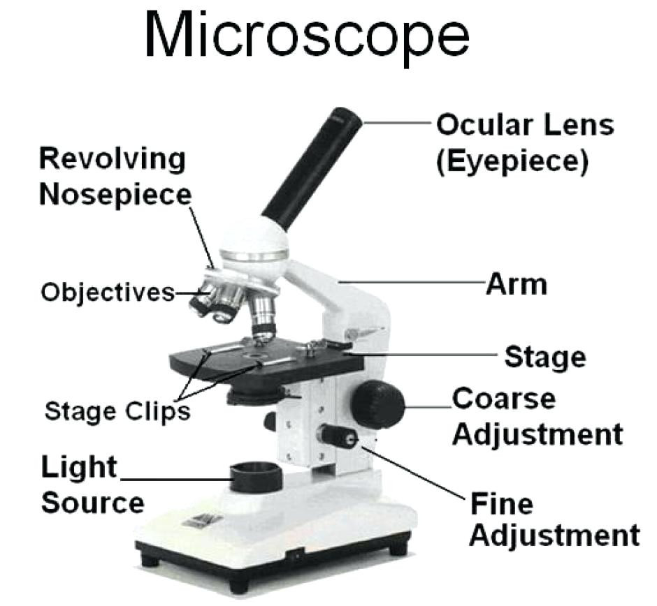
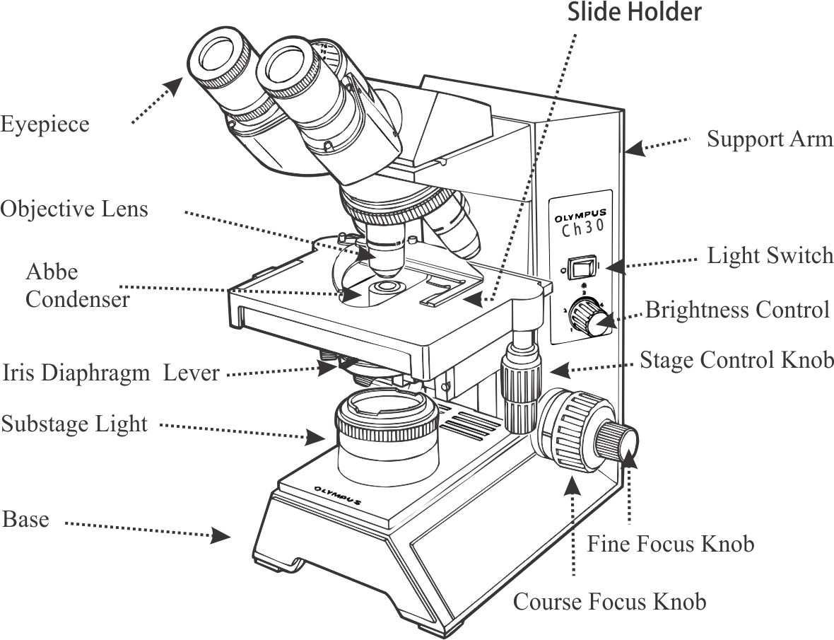
Microscope Diagram Labeled, Unlabeled and Blank | Parts of a Microscope ... This PDF contains the following: 1. Parts of a Microscope Diagram - Color 2. Parts of a Microscope Diagram - Black and White 3. Blank Parts of a Microscope Diagram - Black and White 4. Blank, Unlabeled Parts of a Microscope Diagram - Black and White 5. Blank Parts of a Microscope Diagram - Color 6. Blank, Unlabeled… Label Microscope Diagram - EnchantedLearning.com arm - this attaches the eyepiece and body tube to the base. base - this supports the microscope. body tube - the tube that supports the eyepiece. coarse focus adjustment - a knob that makes large adjustments to the focus. diaphragm - an adjustable opening under the stage, allowing different amounts of light onto the stage. eyepiece - where you place your eye.
Compound Microscope Parts - Labeled Diagram and their Functions - Rs ... The eyepiece (or ocular lens) is the lens part at the top of a microscope that the viewer looks through. The standard eyepiece has a magnification of 10x. You may exchange with an optional eyepiece ranging from 5x - 30x. [In this figure] The structure inside an eyepiece. The current design of the eyepiece is no longer a single convex lens.
Easy microscope diagram with labels
mathsmadeeasy.co.uk › wp-content › uploadsOxford Cambridge and RSA Friday 16 October 2020 – Morning - MME 2 OCR 2020 Answer all the questions. 1 (a) A student was observing onion epithelial cells using a light microscope. They photographed these cells and the image obtained is shown in Fig. 1.1. Simple Microscope - Parts, Functions, Diagram and Labelling Simple Microscope - Parts, Functions, Diagram and Labelling A microscope is one of the commonly used equipment in a laboratory setting. A microscope is an optical instrument used to magnify an image of a tiny object; objects that are not visible to the human eyes. Table of Contents The common types of microscopes are: What is a Simple microscope? Label Microscope Diagram - EnchantedLearning.com arm - this attaches the eyepiece and body tube to the base. base - this supports the microscope. body tube - the tube that supports the eyepiece. coarse focus adjustment - a knob that makes large adjustments to the focus. diaphragm - an adjustable opening under the stage, allowing different amounts of light onto the stage.
Easy microscope diagram with labels. The Parts of a Microscope (Labeled) Printable - TeacherVision This diagram labels and explains the function of each part of a microscope. Use this printable as a handout or transparency to help prepare students for working with laboratory equipment. ... The Parts of a Microscope (Labeled) Printable. Download. Add to Favorites. CREATE NEW FOLDER. Cancel. Manage My Favorites. Share. This diagram labels and ... Microscope Drawing And Label - Painting Valley label microscope diagram compound parts light labeling functions microscopic blank labeled biology microscopy labelled beautiful Compound Microscope ... 496x600 35 0 Parts Of A Compound ... 500x469 27 0 Microscopic Drawing ... 1024x1024 21 4 Download The Diagram... 547x579 17 0 Microscope Labeling ... 270x350 17 0 Microscope Labeling ... › topics › medicine-andConfocal Microscopy - an overview | ScienceDirect Topics John M. Murray, in Methods in Cell Biology, 2013 Abstract. Confocal microscopes are in principle well suited for quantitative imaging. The 3D fluorophore distribution in a specimen is transformed by the microscope optics and detector into the 2D intensity distribution of a digital image by a linear operation, a convolution. Parts of the Microscope with Labeling (also Free Printouts) Parts of the Microscope with Labeling (also Free Printouts) A microscope is one of the invaluable tools in the laboratory setting. It is used to observe things that cannot be seen by the naked eye. Table of Contents 1. Eyepiece 2. Body tube/Head 3. Turret/Nose piece 4. Objective lenses 5. Knobs (fine and coarse) 6. Stage and stage clips 7. Aperture
Microscope Labeling - The Biology Corner 1) Start with scanning (the shortest objective) and only use the COARSE knob . Once it is focused… 2) Switch to low power (medium) and only use the COARSE knob . You may need to recenter your slide. Once it is focused.. 3) Switch to high power (long objective). Parts of a Simple Microscope - Labeled (with diagrams) Parts of a Simple Microscope - Labeled (with diagrams) A simple microscope is a very first type of microscope ever created. It consists of simple parts and performs simple functions. Although there are now many advanced microscope types, some applications may still demand the use of a simple microscope. Microscope Parts Diagram - schemaeasy.com Parts of a microscope with functions and labeled diagram Color the diaphragm light green. Study a drop of pond water to look for microorganisms Collect water samples from a scam or pond bottom. When you are finished using your microscope, turn off the switch, remove the specimen, unplug the power cord, and cover the microscope with its dust cover. Labeling the Parts of the Microscope | Microscope World Resources Labeling the Parts of the Microscope. This activity has been designed for use in homes and schools. Each microscope layout (both blank and the version with answers) are available as PDF downloads. You can view a more in-depth review of each part of the microscope here.
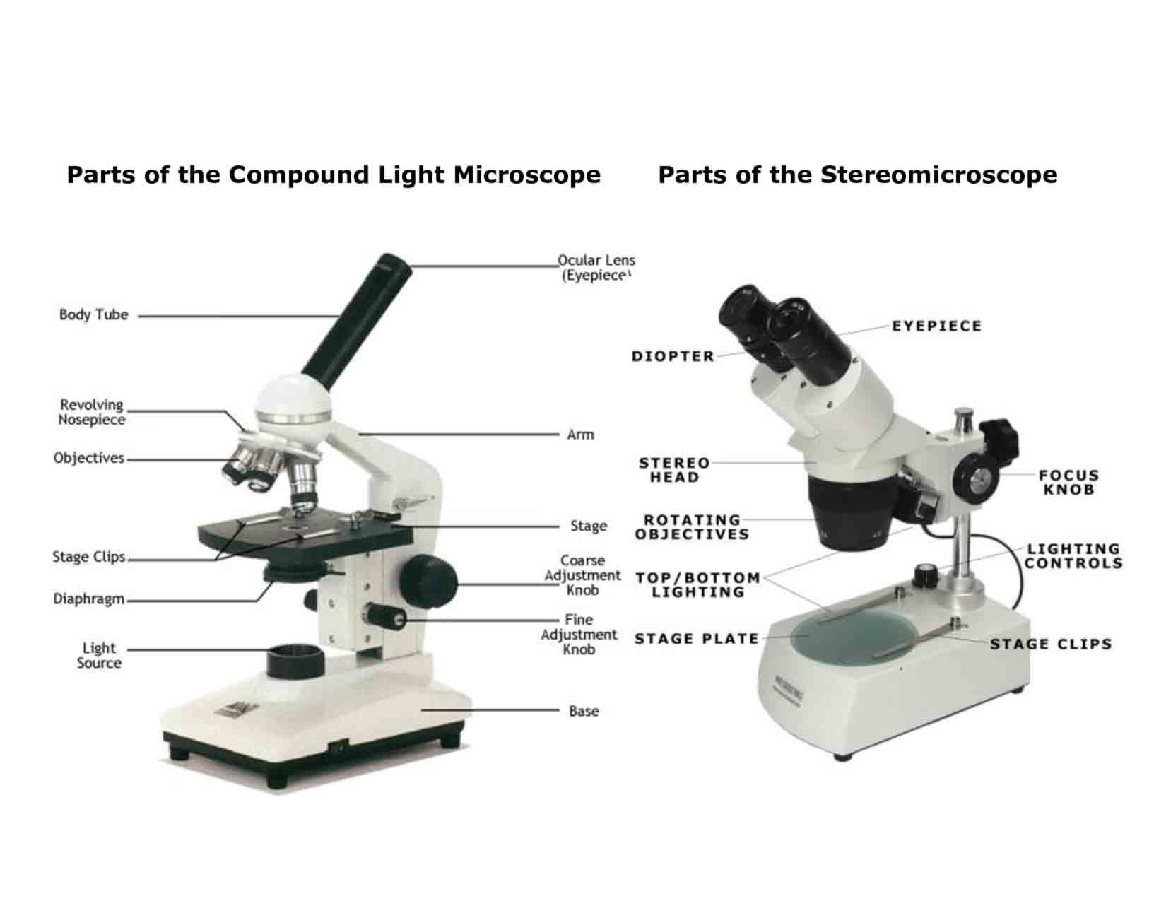
Labelled Diagram of Compound Microscope - Biology Discussion The below mentioned article provides a labelled diagram of compound microscope. Part # 1. The Stand: The stand is made up of a heavy foot which carries a curved inclinable limb or arm bearing the body tube. The foot is generally horse shoe-shaped structure (Fig. 2) which rests on table top or any other surface on which the microscope in kept. A Study of the Microscope and its Functions With a Labeled Diagram ... A Study of the Microscope and its Functions With a Labeled Diagram To better understand the structure and function of a microscope, we need to take a look at the labeled microscope diagrams of the compound and electron microscope. These diagrams clearly explain the functioning of the microscopes along with their respective parts. M mooketsi Parts of a microscope with functions and labeled diagram Figure: Diagram of parts of a microscope There are three structural parts of the microscope i.e. head, base, and arm. Head - This is also known as the body. It carries the optical parts in the upper part of the microscope. Base - It acts as microscopes support. It also carries microscopic illuminators. Label the microscope — Science Learning Hub All microscopes share features in common. In this interactive, you can label the different parts of a microscope. Use this with the Microscope parts activity to help students identify and label the main parts of a microscope and then describe their functions. Drag and drop the text labels onto the microscope diagram.
33 Diagram Of Spirogyra With Label - Labels Design Ideas 2020
Easy labeled diagram of Microscope - YouTube Current diagram shows a detailed diagram of a light microscope which is the most important instrument used in biology and other science fields to observe mic...

This is a quiz called Microscope Labeling Game and was created by member… | Microscope parts ...
Microscope Drawing Easy with Label - YouTube In this video I go over a microscope drawing that is easy with label. There is a blank copy at the end of the video to review on your own. A great way to s...
Labelled Microscope with Functions Storyboard Szerint oliversmith
Simple Microscope - Diagram (Parts labelled), Principle, Formula and Uses Parts of a Simple Microscope A simple microscope consists of Optical parts Mechanical parts Labeled Diagram of simple microscope parts Optical parts The optical parts of a simple microscope include Lens Mirror Eyepiece Lens A simple microscope uses biconvex lens to magnify the image of a specimen under focus.

All Saints Online
› books › NBK26880Looking at the Structure of Cells in the Microscope Many light-microscope techniques are available for observing cells. Cells that have been fixed and stained can be studied in a conventional light microscope, while antibodies coupled to fluorescent dyes can be used to locate specific molecules in cells in a fluorescence microscope. Living cells can be seen with phase-contrast, differential ...

Compound Light Microscope Labeled - Made By Creative Label
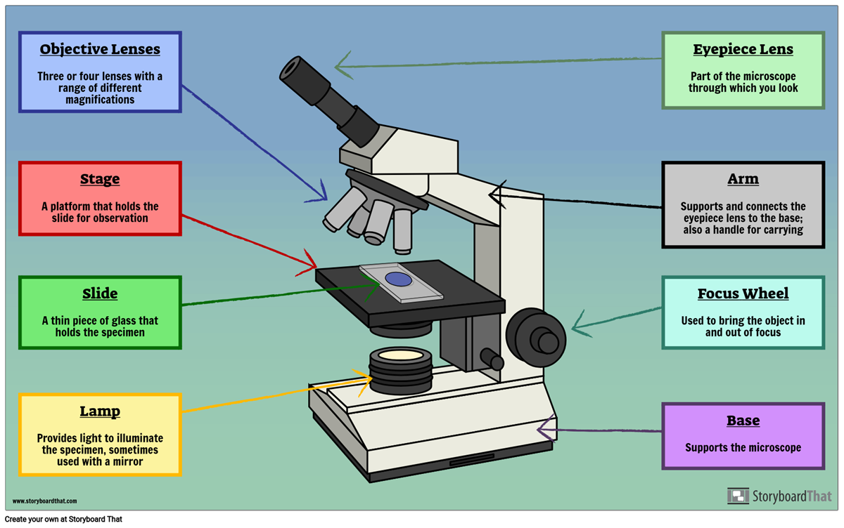
Microscope, Microscope Parts, Labeled Diagram, and Functions Revolving Nosepiece or Turret: Turret is the part of the microscope that holds two or multiple objective lenses and helps to rotate objective lenses and also helps to easily change power. Objective Lenses: Three are 3 or 4 objective lenses on a microscope. The objective lenses almost always consist of 4x, 10x, 40x and 100x powers. The most common eyepiece lens is 10x and when it coupled with ...
Simple Parts Of A Microscope Diagram - Micropedia
› publication › 320945390(PDF) Introduction to Microscopy - ResearchGate 1. Microscopy with light and electrons 2. Electron/specimen interactions: processes and detectors 3. The electron microscope family 4. Specimen preparation for electron microscopy 5.
Simple Parts Of A Microscope Diagram - Micropedia
rsscience.com › stereo-microscopeParts of Stereo Microscope (Dissecting microscope) - Rs' Science Labeled part diagram of a stereo microscope Major structural parts of a stereo microscope. There are three major structural parts of a stereo microscope. The viewing Head includes the upper part of the microscope, which houses the most critical optical components, including the eyepiece, objective lens, and light source of the microscope.
Light Microscope Main Parts Of Light Microscope Biology — db-excel.com
Label A Microscope Worksheets & Teaching Resources | TpT PDF. This packet has 13 pages of awesome resources for introducing and reviewing basic cell anatomy and theory. Perfect for classroom activities or homeschool!Page 1: Mini-poster (8.5"x11" showing organelles in typical plant and animal cells)Page 2: "Blank" animal cell to labelPage 3: "Blank" plant cell.

A School Called Home: A View Through the Microscope
PDF Parts of a Microscope Printables - Homeschool Creations Label the parts of the microscope. You can use the word bank below to fill in the blanks or cut and paste the words at the bottom. Microscope Created by Jolanthe @ HomeschoolCreations.net. Parts of a eyepiece arm stageclips nosepiece focusing knobs illuminator stage objective lenses

labelled diagram of microscope - Brainly.in
› teacher-resources › InteractiveHot and Cold Packs: A Thermochemistry Activity - Carolina.com Diagram your hot or cold pack. Include labels to indicate sizes and quantities of materials used. List all materials and quantities needed to create your thermal pack. Explain the steps that you will follow to build your thermal pack. Describe the safety precautions you will use when creating and testing the thermal pack.

Microscope Labelled Diagram Gcse - Micropedia
› topics › chemistryOptical Sensor - an overview | ScienceDirect Topics A far-field, epifluorescence microscope system for single tube spectroscopy was first proposed by Weisman and co-workers, and its schematic diagram is presented in Fig. 10.11. 23, 27 Visible images of sample morphology can be viewed through the eyepiece of the microscope or through a CCD camera. The samples are photo-excited by lasers, and ...
template
Microscope Poster - Diagram with Labels | Teach Starter A poster containing a diagram with labels showing the key parts of a microscope. In Science it is important that students know how to use a variety of tools when conducting scientific experiments and inquiry. This poster focuses on the microscope and highlights its key parts. There are two print options available for this poster:

Dense Regular Connective Tissue Labeled - Made By Creative Label
Labeling Microscope Worksheet | Teaching Resources A straightforward worksheet in which students are required to identify the parts of a basic microscope. Tes classic free licence. Report this resource to let us know if it violates our terms and conditions. Our customer service team will review your report and will be in touch. Last updated. 21 November 2014.
Post a Comment for "43 easy microscope diagram with labels"