39 diagram of a human cell with labels
Diagram of human skin structure — Science Learning Hub Diagram of human skin structure. Add to collection. + Create new collection. Tweet. Rights: University of Waikato Published 1 February 2011 Size: 100 KB Referencing Hub media. The epidermis is a tough coating formed from overlapping layers of dead skin cells. Label Diagram Human Body Stock Illustrations - 161 Label ... Download 161 Label Diagram Human Body Stock Illustrations, Vectors & Clipart for FREE or amazingly low rates! New users enjoy 60% OFF. 185,925,055 stock photos online. ... Animal cell structure anatomy infographic diagram. With parts flat vector illustration design for biology science education school book concept microbiology.
Anatomy (Human Body) Labeling - Exploring Nature Muscles of the Leg and Foot Labeling Page. Muscles of the Neck, Chest and Thorax Labeling Page. Muscles of the Neck, Shoulders and Thorax (Posterior) Labeling. Muscles of the Posterior Body Labeling (HS-Adult) Muscles of the Thigh and Hip (Anterior) Labeling. Muscles of the Thigh and Hip (Posterior) Labeling. Nerve Cell (Neuron) Labeling Page.

Diagram of a human cell with labels
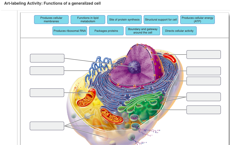
Thyroid gland: cells, tissues, labeled diagram (preview ... This is a sneak peek at the full tutorial about the thyroid gland histology. Watch the full video at Kenhub: , are you struggling with... Animal Cell Diagram | Science Trends An animal cell diagram is a great way to learn and understand the many functions of an animal cell. The diagram, like the one above, will include labels of the major parts of an animal cell including the cell membrane, nucleus, ribosomes, mitochondria, vesicles, and cytosol. Animal Cells: Labelled Diagram, Definitions, and Structure Animal Cells Organelles and Functions. A double layer that supports and protects the cell. Allows materials in and out. The control center of the cell. Nucleus contains majority of cell's the DNA. Popularly known as the "Powerhouse". Breaks down food to produce energy in the form of ATP.
Diagram of a human cell with labels. Labeled Plant Cell With Diagrams | Science Trends The parts of a plant cell include the cell wall, the cell membrane, the cytoskeleton or cytoplasm, the nucleus, the Golgi body, the mitochondria, the peroxisome's, the vacuoles, ribosomes, and the endoplasmic reticulum. Parts Of A Plant Cell The Cell Wall Let's start from the outside and work our way inwards. Cell: Structure and Functions (With Diagram) Eukaryotic Cells: 1. Eukaryotes are sophisticated cells with a well defined nucleus and cell organelles. 2. The cells are comparatively larger in size (10-100 μm). 3. Unicellular to multicellular in nature and evolved ~1 billion years ago. 4. The cell membrane is semipermeable and flexible. 5. These cells reproduce both asexually and sexually. Animal Cell Diagram High Resolution Stock Photography and ... Find the perfect animal cell diagram stock photo. Huge collection, amazing choice, 100+ million high quality, affordable RF and RM images. ... The structure of a human's cell with labeled parts. cross section of a Eukaryotic cell. ... Internal Diagram Structure of Human Cell on a white background. 3d Rendering Fission Simple vector icon. Human ... Pin by Lahu deore on Dc. | Human cell diagram, Cell ... This diagram of a human skeleton is labeled with 12 major bones, from skull to fibula. D Nicole Science Tissue Biology Anatomy And Physiology Textbook Histology Slides Biology College Basic Physics Chemistry Lessons Nursing School Notes 6th Grade Science Medical Coding 18" by 24" Laminated Wall Chart A My Airtel Nursing
Cell Membrane Diagram Labeled : Functions and Diagram Cell Membrane Diagram. There are no organelles in the prokaryotic cells, i.e., they have no internal membrane systems. While lipids help to give membranes their flexibility, proteins monitor and maintain. We all keep in mind that the human body is very elaborate and a technique I found out to understand it is by means of the manner of human ... Labeled Diagram of the Human Kidney - Bodytomy Labeled Diagram of the Human Kidney The human kidneys house millions of tiny filtration units called nephrons, which enable our body to retain the vital nutrients, and excrete the unwanted or excess molecules as well as metabolic wastes from the body. In addition, they also play an important role in maintaining the water balance of our body. PDF Human Cell Diagram, Parts, Pictures, Structure and Functions Diagram of the human cell illustrating the different parts of the cell. Cell Membrane The cell membraneis the outer coating of the cell and contains the cytoplasm, substances within it and the organelle. It is a double-layered membrane composed of proteins and lipids. Circulatory System Labeled Diagram Illustrations, Royalty ... Browse 154 circulatory system labeled diagram stock illustrations and vector graphics available royalty-free, or start a new search to explore more great stock images and vector art. Newest results Heart Poster Heart blood flow circulation anatomical diagram with atrium and... Anatomy of Nerves of Body and Head
Karyotype Diagram - SmartDraw Karyotype. A karyotype is a complete set of all chromosomes of a cell of any living organism. Karyotypes are examined in searches for chromosomal aberrations such as genetic disorders, and can also be used to determine other macroscopically visible aspects such as gender. Normal Male Karyotype. Normal Female Karyotype. The Human Skeleton: All You Need to Know - Bodytomy Labeled Skeleton Diagram This skeleton diagram will help explain the different bones of the human body clearly. Cranium The cranium is a skull bone that covers the brain, as seen in the skeleton diagram. The facial bones are not a part of the cranium. The bones that are just above the ear or in front of the ear are known as temporal bones. Stapes Animal Cell Diagram with Label and Explanation: Cell ... Below is the diagram of the animal cell which shows the organelles present in it. The cell is covered with cytoplasm which consists of cell organelles in it. The nucleus is covered with a rough Endoplasmic Reticulum and other organelles each designed for a specific purpose. A Labeled Diagram of the Animal Cell and its Organelles ... As observed in the labeled animal cell diagram, the cell membrane forms the confining factor of the cell, that is it envelopes the cell constituents together and gives the cell its shape, form, and existence. ... it is essential that the DNA remains intact and gets evenly distributed among the cells. Every human body cell contains 46 ...

Explain the nucleus of a cell with a neat labeled diagram - Science - Cell - Structure and ...
What Is Going On Inside That Cell? | Human cell diagram ... cell, in biology, the basic membrane-bound unit that contains the fundamental molecules of life and of which all living things are composed. A single cell is often a complete organism in itself, such as a bacterium or yeast. Other cells acquire specialized functions as they mature.
Skeletal System - Labeled Diagrams of the Human Skeleton The skeletal system's cell matrix acts as our calcium bank by storing and releasing calcium ions into the blood as needed. Proper levels of calcium ions in the blood are essential to the proper function of the nervous and muscular systems. Bone cells also release osteocalcin, a hormone that helps regulate blood sugar and fat deposition.
Skin Diagram with Detailed Illustrations and Clear Labels Skin Diagram with Detailed Illustrations and Clear Labels Biology Important Diagrams Skin Diagram Skin Diagram The largest organ in the human body is the skin, covering a total area of about 1.8 square meters. The skin is tasked with protecting our body from the external elements as well as microbes. Interesting Note:
Human Cell - TheInspiredInstructor.com DIRECTIONS: Type the number beside the name of the cell part into the box before its description. (1) A thin covering which encloses the cell and regulates substances that pass through it. (2) A jelly-like material which fills the cell (3) The control center of a cell which directs its activities.
Heart Diagram with Labels and Detailed Explanation Diagram of Heart. The human heart is the most crucial organ of the human body. It pumps blood from the heart to different parts of the body and back to the heart. The most common heart attack symptoms or warning signs are chest pain, breathlessness, nausea, sweating etc. The diagram of heart is beneficial for Class 10 and 12 and is frequently ...
03 Label the Cell Diagram | Quizlet Start studying 03 Label the Cell. Learn vocabulary, terms, and more with flashcards, games, and other study tools.
Label the cell diagram - Teaching resources Label the Plot Diagram - Label the Cell Membrane - Label the Cell Membrane - Label the Plant Cell - Label the Cell - Label the Cell Cycle - Label the Plant Cell ... The diagram shows various endocrine glands in human body. Label the diagram. Labelled diagram. by Deeksha7. Animal Cell Diagram Labeling Labelled diagram. by Brittney7. G7 Biology ...
Blood Cell Diagram Stock Photos, Pictures & Royalty-Free ... Bone marrow Blood stem cell is an immature cell that can develop into all types of blood cells, including white blood cells, red blood cells, and platelets. Blood stem cells are found in the peripheral blood and the bone marrow. Also called hematopoietic stem cell. 3d render blood cell diagram stock pictures, royalty-free photos & images.

Questions And Answers On Labeled/Unlebled Diagrams Of A Human Cell : Questions And Answers On ...
Human Cell Diagram, Parts, Pictures, Structure and ... One of the few cells in the human body that lacks almost all organelles are the red blood cells. The main organelles are as follows : cell membrane endoplasmic reticulum Golgi apparatus lysosomes mitochondria nucleus perioxisomes microfilaments and microtubules Diagram of the human cell illustrating the different parts of the cell. Cell Membrane
Animal Cells: Labelled Diagram, Definitions, and Structure Animal Cells Organelles and Functions. A double layer that supports and protects the cell. Allows materials in and out. The control center of the cell. Nucleus contains majority of cell's the DNA. Popularly known as the "Powerhouse". Breaks down food to produce energy in the form of ATP.
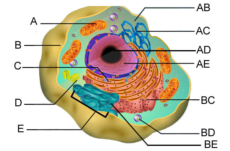
Questions And Answers On Labeled/Unlebled Diagrams Of A Human Cell / Questions And Answers On ...
Animal Cell Diagram | Science Trends An animal cell diagram is a great way to learn and understand the many functions of an animal cell. The diagram, like the one above, will include labels of the major parts of an animal cell including the cell membrane, nucleus, ribosomes, mitochondria, vesicles, and cytosol.
Thyroid gland: cells, tissues, labeled diagram (preview ... This is a sneak peek at the full tutorial about the thyroid gland histology. Watch the full video at Kenhub: , are you struggling with...


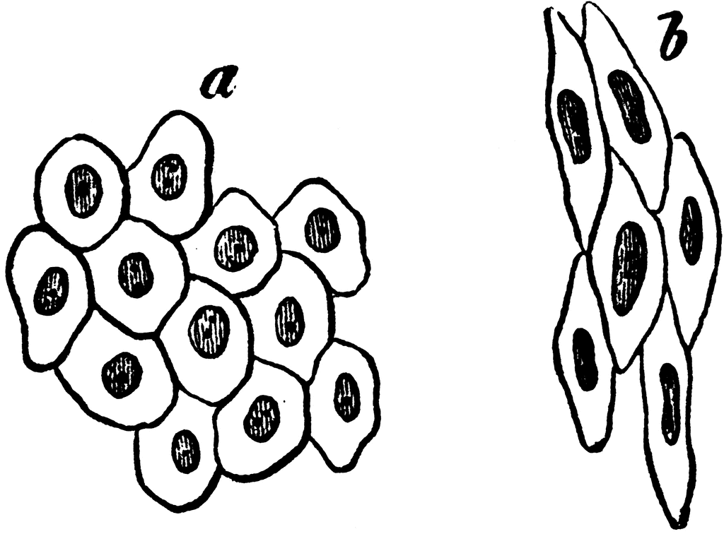

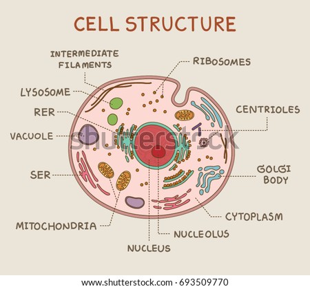
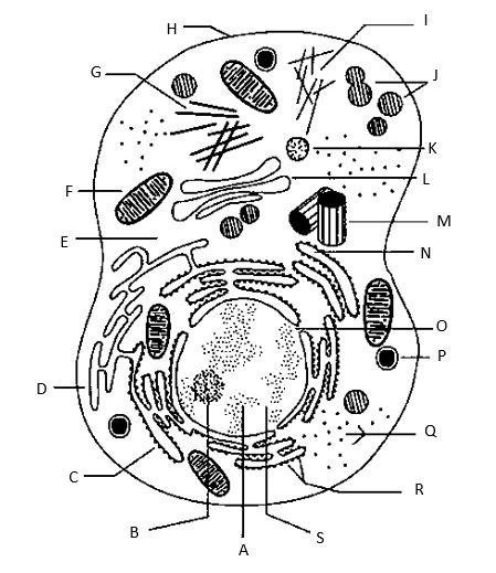


Post a Comment for "39 diagram of a human cell with labels"